
Dermatoscopy & Mole Imaging
Enhance early detection with advanced dermatoscopy and mole imaging services at Dermadocs Skin Cancer Clinic in Parkside, Adelaide. These specialised technologies help clinicians examine, document and monitor skin lesions and moles with precision — improving diagnostic accuracy and supporting long-term skin surveillance.
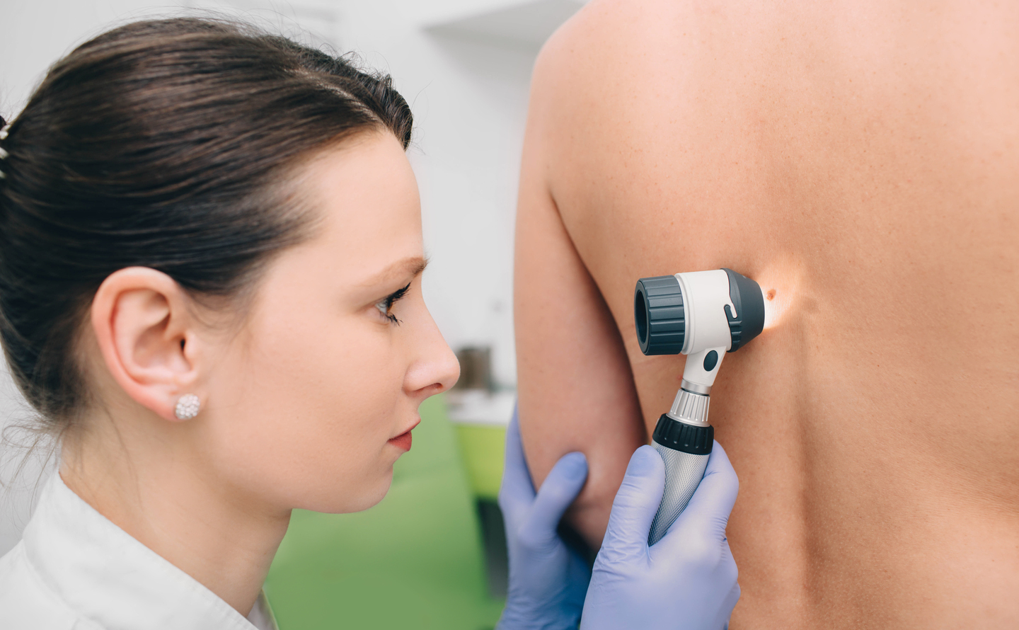
Dermatoscopy (also called dermoscopy or epiluminescence microscopy) is a non-invasive visual examination of skin lesions using a handheld or digital imaging device known as a dermatoscope. This device combines a magnifier and specialised lighting to show structures and patterns beneath the skin surface that are not visible to the naked eye, aiding in early and accurate detection of melanoma and other skin cancers.
Dermatoscopy significantly increases diagnostic precision compared with a standard visual exam, helping clinicians identify subtle features of pigmented and non-pigmented lesions.
Dermatoscopy & Mole Imaging is especially valuable if you: • Have a personal or family history of melanoma or skin cancer • Have numerous moles or atypical spots • Want structured long-term surveillance • Are at higher risk because of lighter skin or extensive UV exposure
A complete visual examination of your skin from head to toe under professional lighting conditions, looking for new or changing spots, moles or lesions. This forms the foundation of early detection.
Focused evaluation of one or more specific areas of concern, recommended if you’ve noticed changes or symptoms in a particular spot.
Photographic mapping of the entire skin surface to document and monitor numerous moles or lesions, especially for patients at high risk. It supports detailed comparison over time and helps reveal changes that may require intervention.
During your skin check, your clinician will examine areas of concern visually and with a dermatoscope.
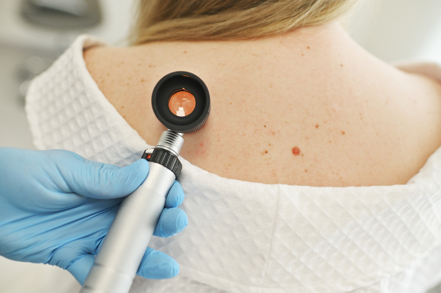
If needed, digital photos are taken of individual lesions or moles for documentation.
Images are reviewed and stored securely, forming a reference set that supports future comparisons.
Based on findings, your clinician will recommend a monitoring frequency, which may include routine re-imaging on subsequent visits.
Dermatoscopy uses a specialised magnifying device to examine skin lesions more closely, offering greater detail than a standard visual check.
No — imaging is non-invasive, quick and painless, involving only digital photography of the skin surface.
Yes — stored images can be used to track changes over time and assess mole evolution between visits.